

In This Article
If you’re considering veneers while bruxism is suspected, it’s more reasonable to assess factors that increase the risk of damage rather than drawing a conclusion from a single test. In this context, an in-office EMG screening can be a helpful reference because it quantifies muscle-use patterns when you clench your teeth and helps organize potential warning signals. This article summarizes the basic workflow of bruxism diagnosis, the supportive role of EMG testing, and systematic management criteria to reduce the risk of restoration damage.
1. Why doesn’t bruxism diagnosis end with a single test?
Rather than confirming bruxism with a single device-based test, it is more appropriate to interpret the condition by integrating the patient’s subjective symptoms, a clinical oral examination, and—when needed—instrument-based assessments. Because sleep-related oral habits are often outside a person’s awareness, the first step is establishing when and under what circumstances bruxism should be suspected.
Clues such as a bed partner’s observations, jaw fatigue upon waking, and frequent breakage of restorations are important elements of the medical history. In contrast, tooth marks on the tongue or inner cheeks, hypertrophy of the chewing muscles, and microcracks on tooth surfaces are objective clinical findings that can be confirmed in the dental office.
Only after these pieces of information are gathered can the clinician clearly determine at what stage an instrument-based assessment such as EMG testing will be useful.
2. What can an EMG test tell us?
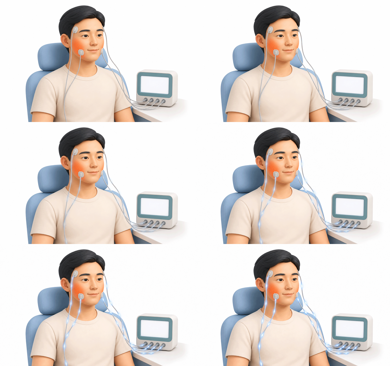
An in-office surface electromyography (sEMG) screening is useful for numerically assessing how strongly the muscles tense when you clench your teeth and how symmetric left–right muscle use is. In particular, it provides baseline data to identify whether someone shows an overactivation pattern and to decide whether additional safety measures should be included in the treatment plan.
However, it is difficult to treat a short clenching measurement as perfectly equivalent to indicators of the frequency or intensity of sleep bruxism.
If snoring, suspected sleep apnea, or daytime sleepiness is present, a separate overnight evaluation such as polysomnography may be considered. In that case, EMG values are best understood as supportive indicators that further clarify potential risk signals.
3. Key conservative criteria to reduce the risk of veneer fracture
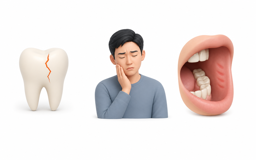
Having bruxism does not automatically mean veneers will fracture, but strong chewing forces can be a major risk factor for restoration failure. To withstand heavy loads, it is not enough to simply choose a strong ceramic material; the case should also meet minimally invasive preparation and bonding criteria that preserve as much natural enamel as possible.
As the exposed dentin area increases, bonding strength may decrease, which can negatively affect long-term survival. Therefore, it is important to closely review any history of restoration fracture and the occlusal scheme, while also incorporating a design that disperses load concentrations.
✅ Key clues that may increase risk
- A history of repeated debonding of existing restorations or a diagnosis of tooth cracks
- Frequent morning jaw fatigue and noticeably enlarged masseter (chewing) muscles
- Recurrent tooth-pressed marks (indentations) on the tongue and inner cheek mucosa
4. If there is a lot of tooth wear, does that mean it’s severe right now?
Tooth wear can be a nonspecific trace accumulated over time, so it is difficult to conclude that current sleep bruxism activity is severe based on wear alone. Worn tooth surfaces may remain like old tire tracks, but they do not directly show the recent intensity of friction or how often it has occurred.
For this reason, clinical care places more weight on recent signals of change in the oral environment than on older traces. A noticeable increase in tooth cracks lately, or more frequent mornings with jaw soreness, can serve as much more direct clues.
When needed, an EMG screening can be used to check current muscle tension, and objective indicators can be narrowed step by step so patients do not become overly anxious about past signs.
5. Botox and a night guard: do I need them too?
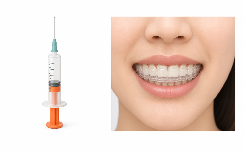
Botox treatment and a night guard do not serve the same purpose; they are options that protect the teeth through different mechanisms. Botox may help temporarily reduce the strength of chewing-muscle contraction or the amplitude of muscle activity, but it should be approached separately from a cause-targeting treatment that eliminates the occurrence of bruxism itself.
A night guard, on the other hand, focuses on mechanically covering and protecting the teeth and restorations by evenly distributing physical loads during sleep. It is advisable to decide—together with a clinician—whether to combine these approaches after a comprehensive evaluation of fracture history, the amount of tooth reduction, and the degree of enamel preservation.
6. Frequently Asked Questions (FAQ)
Q. If my surface EMG (sEMG) value is high, does that mean I can’t receive veneer treatment?
In-office sEMG is a supportive indicator that quantifies how much the muscles tense when you clench, so treatment eligibility is not determined by a single number. Clinicians can help you proceed safely after considering your history of restoration fracture, current occlusal design, bonding conditions, and whether protective devices will be used.
Q. Do I need polysomnography to manage bruxism?
Polysomnography is not required for everyone with suspected bruxism. A detailed overnight evaluation is typically discussed for patients in whom signs of breathing-related sleep disorders—such as significant snoring during sleep, suspected sleep apnea, and daytime sleepiness—are observed together.
Q. What does a night guard do, and how often should it be checked?
A night guard acts as a protective barrier by evenly distributing strong friction and loads that occur unconsciously. With long-term use, the surface may wear down or the bite may change subtly, so it is safer to receive regular occlusal checkups according to the schedule provided by your clinician.
Q. Does Botox eliminate bruxism at its source?
Botox therapy may play a supportive role by weakening the contraction force of the chewing muscles and reducing the burden on the jaw and restorations. Because it does not remove the triggers that cause bruxism, it is appropriate to discuss in detail with a clinician whether to combine it with other protective strategies such as an occlusal stabilization appliance.
This health content has been reviewed by medical professionals, compiled from authoritative domestic and international sources, and is regularly updated in accordance with the latest clinical guidelines.
Sources
- Korea Disease Control and Prevention Agency (KDCA) National Health Information Portal, Clinical diagnostic guidelines for bruxism and criteria for using supportive device-based tests, 2024.
- Al‑Talib et al., Bruxism and direct and indirect restorations failure: A scoping review, Journal of Dentistry, 2025.
- Nguyen et al., Coexisting pain‑related TMD and probable sleep bruxism, BMC Oral Health, 2026.
※ The copyright for all content on this blog belongs to medihi. Unauthorized copying, distribution, or derivative use is strictly prohibited, and violations may result in legal action without prior notice.