

In This Article
In middle age, pain behind the knee and chronic swelling can make walking uncomfortable. If you notice a lump behind the knee and a tight, “stuffed” sensation when bending, it’s important to evaluate not only the lump itself but also the condition inside the joint. This article explains why a Baker’s cyst develops, how to differentiate calf swelling, the roles of ultrasound and MRI, and symptom-based criteria for treatment and ongoing management.
1. Why does a Baker’s cyst develop?
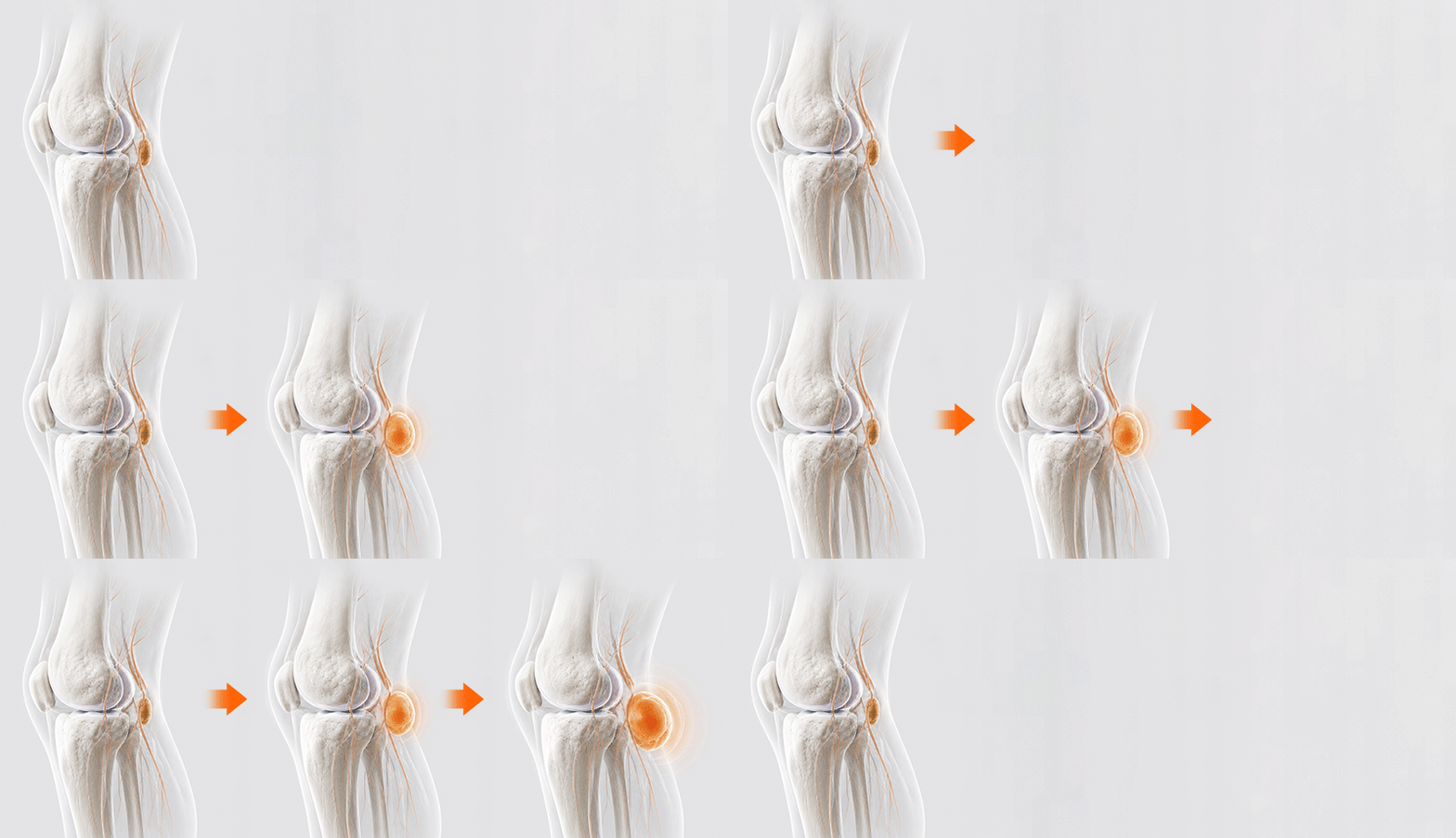
A Baker’s cyst is a condition in which excessive knee joint fluid is produced and pushed backward, where it collects; it is also called a popliteal cyst. In adults, a Baker’s cyst is often less a standalone problem and more a sign that joint fluid has increased abnormally due to cartilage or ligament issues. It is frequently found along with intra-articular problems such as knee osteoarthritis or a meniscal tear.
When inflammation occurs in the joint, synovial fluid (which acts like a lubricant) increases. This fluid is forced backward through a one-way, valve-like mechanism that prevents it from flowing back into the joint, forming a sac.
In treatment, it is more important to evaluate the underlying intra-articular cause that is triggering synovitis than to focus only on removing the visible lump. It’s similar to a roof leak: emptying the bucket alone doesn’t fix the leak.
2. If swelling extends into the calf, what should you check?
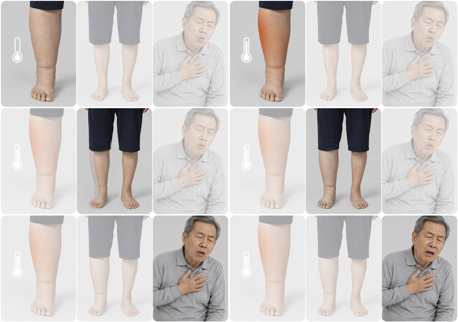
If the calf suddenly becomes very swollen and warm, it is important to distinguish between a ruptured cyst and deep vein thrombosis (DVT). When a cyst ruptures, joint fluid can track down into the muscles and trigger inflammation. Because the symptoms can resemble DVT (a blocked leg vein), it is sometimes called “pseudo-DVT” (also described as pseudothrombophlebitis).
If you mainly feel aching or tightness behind the knee, a joint-related issue is more likely. However, if the calf becomes markedly swollen and red, or if one leg is noticeably more swollen than the other, checking for a clot should come first.
In particular, if chest pain or sudden shortness of breath occurs as well, prompt evaluation is needed. When symptoms are unclear, a medical facility should confirm whether a clot is present using duplex ultrasound (a vascular ultrasound test).
3. When are ultrasound and MRI used?
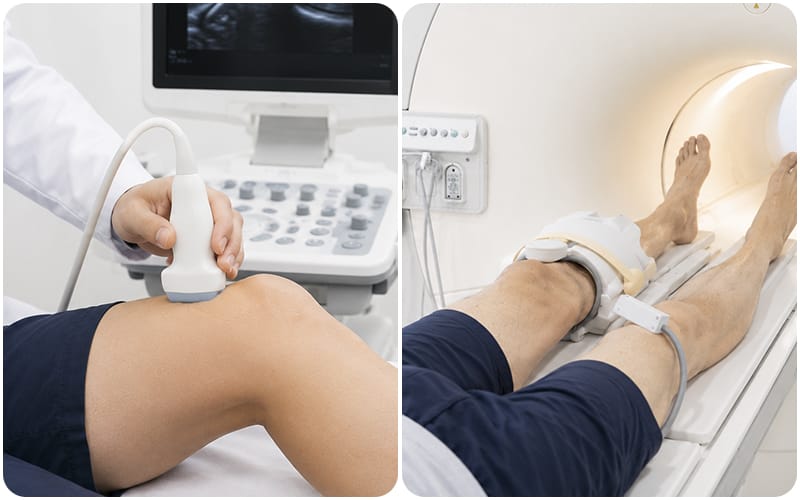
Ultrasound is used to confirm whether a cyst is present and to measure its size, while MRI is more helpful for assessing cartilage and ligament damage inside the joint. The choice of test depends on symptom location and the suspected underlying condition, and not every patient needs advanced imaging from the start.
To confirm the cyst itself, ultrasound is used as the first-line test. It allows real-time assessment of the cyst’s location and size and the amount of joint fluid, and it is also used to guide injection procedures. High diagnostic accuracy has been reported for ultrasound in cyst evaluation.
If symptoms do not improve with conservative treatment, or if the cause needs closer evaluation (such as a meniscal or ligament injury), MRI may be additionally considered.
4. When is fluid drainage or surgery appropriate?
Treatment for a cyst is determined based on its size, pain, pressure symptoms, and whether it recurs repeatedly, while also managing the underlying condition. If it is small and painless, monitoring over time while managing arthritis may be sufficient.
If the tight, “stuffed” sensation is significant, an approach that combines fluid drainage (aspiration) with treatment of the underlying condition may be used. Removing joint fluid can reduce pressure and help with short-term symptom relief. However, if the cause of inflammation inside the joint remains, fluid may reaccumulate within weeks.
If recurrence is frequent or associated conditions are severe, selective surgical decompression to reduce pressure may be considered. Treatment should aim to lower the chance of recurrence, but recovery time and potential complications must also be weighed through thorough discussion with the medical team.
✅Symptoms to check before a consultation
- Check whether pressure behind the knee when bending interferes with daily life.
- Watch whether swelling and pain return within 1–2 months after fluid drainage.
- Check whether the inner side of the knee feels stiff when walking or climbing stairs.
5. Swelling management criteria for women in their 50s–60s
When evaluating chronic swelling in women in their 50s–60s, it is important to assess both knee symptoms and the pattern of leg swelling. Degenerative osteoarthritis and chronic swelling often overlap, and long-term housework or postures that load the knees can also contribute.
If both legs swell together, swelling worsens in the afternoon, and an indentation remains for a long time after pressing (pitting), other causes such as venous circulation issues may be involved. Not all leg swelling is caused by a Baker’s cyst.
Rather than focusing only on reducing the lump size, the priority should be slowing overall degenerative changes. Management should include strengthening the thigh muscles and reducing body weight to decrease joint pressure.
6. Frequently Asked Questions (FAQ)
Q. Is a Baker’s cyst related to knee arthritis?
It is a condition in which excessive joint fluid is produced due to cartilage damage or synovitis and then collects behind the knee. In adults, it is often found together with knee osteoarthritis or a meniscal tear.
Q. If my calf suddenly swells and hurts, how can I tell it apart from a blood clot?
Swelling and warmth after a cyst rupture can look very similar to deep vein thrombosis. Because it is difficult to distinguish based on symptoms alone, vascular ultrasound is needed to confirm whether a clot is present.
Q. Can ultrasound alone diagnose it? When is MRI needed?
Ultrasound can evaluate the cyst’s location and size and the amount of joint fluid. MRI may be additionally needed to identify underlying causes such as cartilage or ligament injuries deeper within the joint.
Q. With what symptoms should I seek care sooner?
If there is no pain and daily life is not affected, you may monitor over time. If pain is severe enough that bending the knee is difficult, or if the entire calf suddenly becomes swollen and warm, it is advisable to first check the underlying cause.
This health content has been reviewed by medical professionals, compiled from authoritative domestic and international sources, and is regularly updated in accordance with the latest clinical guidelines.
Sources
- Korea Ministry of Health and Welfare, National Health Information Portal: Health information on popliteal cyst (Baker’s cyst).
- Korean Orthopaedic Association: Public health information — knee joint disorders.
- Liu et al., Diagnostic accuracy of ultrasound for the assessment of Baker’s cysts: a meta-analysis, 2022.
※ The copyright for all content on this blog belongs to medihi. Unauthorized copying, distribution, or derivative use is strictly prohibited, and violations may result in legal action without prior notice.