

In This Article
When looking for the cause of knee pain, you don’t always need to start with an advanced test like an MRI. The role of X-ray, MRI, and ultrasound differs depending on your symptoms and physical exam findings. Understanding the typical testing sequence can help reduce unnecessary tests and out-of-pocket costs.
1. Knee pain: Which test comes first?

Knee pain is evaluated by first reviewing your medical history and performing a physical exam, then selecting imaging tests step by step as needed. If chronic degenerative changes are suspected in middle-aged or older adults, a weight-bearing X-ray (taken while standing) may be used as the first test.
A weight-bearing X-ray checks bone alignment and joint space while you are standing. It’s used to assess the overall situation—such as whether the joint space has narrowed due to cartilage wear, or whether the leg axis is misaligned. In some cases, this information alone helps identify the cause of degenerative knee pain, meaning an MRI may not be necessary from the start.
Even when knee pain follows a sudden injury, the order of tests is determined based on the exam. If the knee is swollen and it’s difficult to bear weight, an X-ray may be performed first to check for bone injury such as a fracture. If the cause still can’t be explained by basic tests and a ligament or meniscus injury is suspected, an MRI may then be considered.
2. If the X-ray is normal, do you still need an MRI?
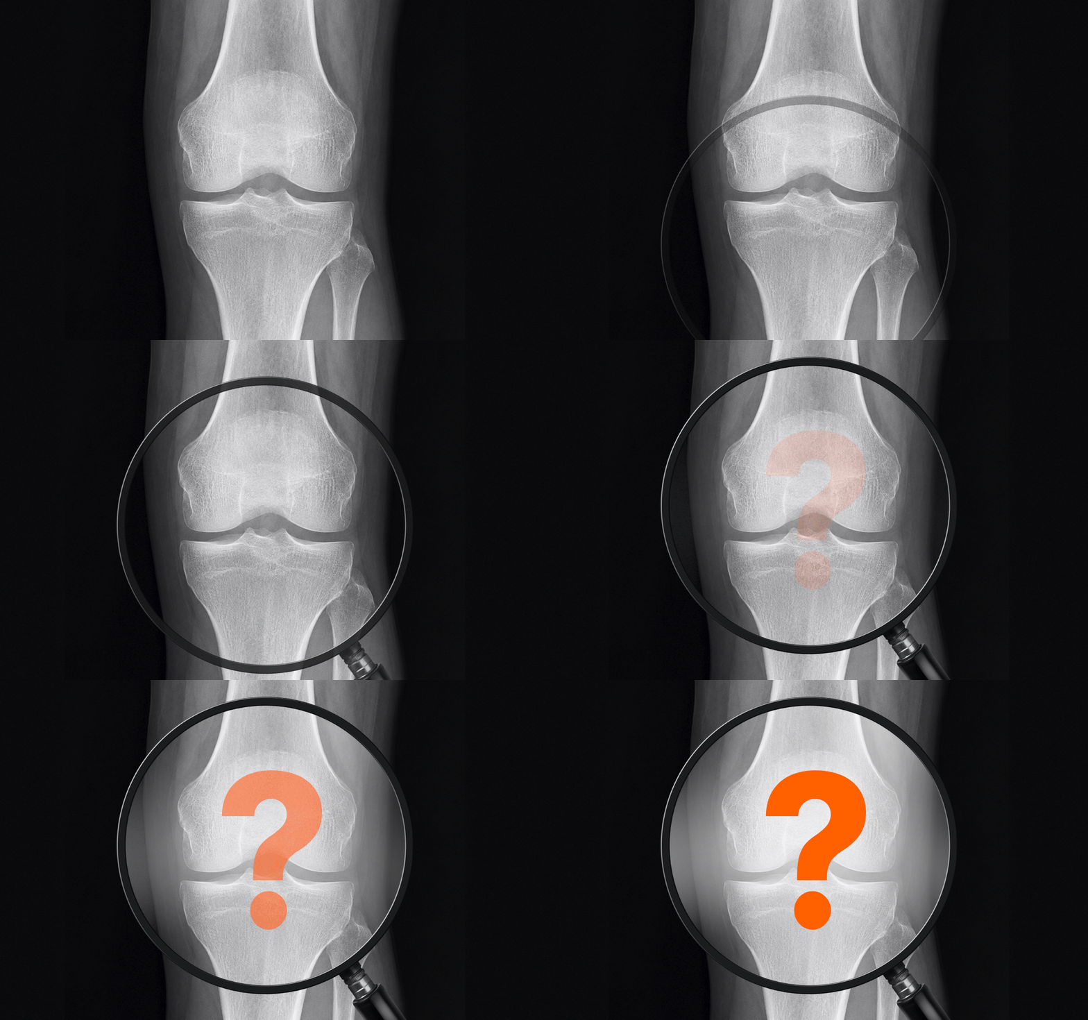
Even if an X-ray appears normal, it does not rule out all causes of pain. X-rays mainly show bone contours, alignment, and joint space, so it’s difficult to directly evaluate soft tissues such as ligaments or the meniscus.
MRI is used when a closer look at these internal structures is needed. Examples include pain that persists for more than 4–6 weeks without a clear explanation from basic testing, or a pronounced “locking” symptom where the knee won’t fully straighten and feels stuck. MRI may also be considered when repeated instability occurs (the knee “gives way” during weight-bearing) or when meniscus or cruciate ligament injury is strongly suspected.
Test selection depends on what needs to be confirmed. A common approach is to first review the overall bony status with X-ray, and then use MRI to evaluate deeper internal tissues when necessary.
3. What are the National Health Insurance coverage criteria for knee MRI?
National Health Insurance coverage for knee MRI is not the same for every type of pain. Coverage is limited, and in particular, chronic degenerative changes that progress gradually without a specific injury—often seen in people in their 40s to 60s—may be more likely to be handled as non-covered (out-of-pocket).
If the situation is closer to degenerative change, such as a chronic meniscus injury, it may be excluded from coverage. On the other hand, coverage may differ depending on whether there was a clear timing of injury and a specific traumatic event such as a fall or twisting incident.
Before your appointment, it can help to organize when the pain or injury began and what situation triggered it. Based on that information, you can review with your clinician whether National Health Insurance applies and what the expected cost range may be.
4. If your knee is swollen and feels like there’s fluid, do you need an ultrasound?
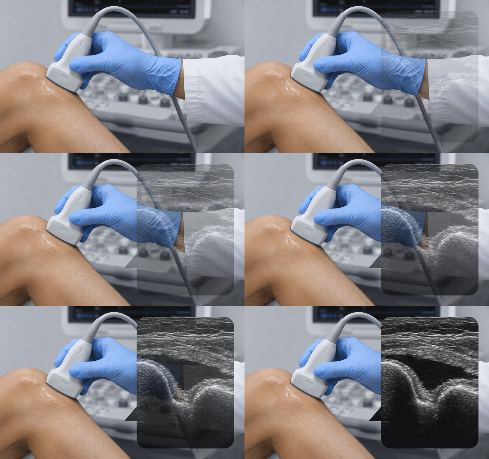
If the knee is significantly swollen or feels like there is fluid in the joint, ultrasound may be used. Ultrasound is useful for real-time evaluation of joint fluid, inflammation near the surface, and tendon condition.
Ultrasound can also be used to guide procedures such as aspirating (draining) joint fluid or performing injection treatment while visualizing the inflamed area. However, it has limitations in closely evaluating deep structures such as the full cruciate ligaments or the internal cartilage.
For that reason, ultrasound can be understood as a test that helps confirm swelling, joint fluid, and more superficial issues—and supports necessary procedures. If you have prior imaging or radiology reports, bringing them to your visit can also be helpful.
5. Can you determine the cause based on imaging results alone?
Even if imaging shows an abnormal finding, it’s difficult to confirm the cause of pain based on that result alone. In middle-aged and older adults, age-related changes such as a meniscus tear may be found incidentally, and these findings do not always match a person’s actual symptoms.
Clinicians make a comprehensive judgment by combining imaging results with the real-world picture—pain severity, swelling, and limitations in knee movement. Even if imaging shows changes, conservative management may be considered if there is no major difficulty with everyday walking.
On the other hand, when imaging findings and actual movement limitations appear together, the information becomes more meaningful for deciding a treatment direction. Knee imaging guidelines are less about a single test result and more about evaluating symptoms, physical exam findings, and imaging together.
6. Frequently Asked Questions (FAQ)
Q. Why is an X-ray taken first?
It’s used to check bone alignment and joint space under weight-bearing conditions. When degenerative changes are suspected, it can serve as a basic test to understand the overall status first.
Q. How does National Health Insurance coverage for MRI differ with a meniscus injury?
It may vary depending on the cause and pattern of injury. Acute injuries with a clear traumatic event and chronic or degenerative-type injuries may differ in whether National Health Insurance coverage applies, so confirmation during the visit is needed.
Q. Can ultrasound evaluate everything inside the knee?
Ultrasound is useful for assessing joint fluid and tissues closer to the surface, but it has limitations in evaluating deep structures like the cruciate ligaments or the full internal cartilage. If needed, other imaging tests may be reviewed together.
Q. What symptoms suggest you need a detailed evaluation?
If the knee won’t straighten or has a catching/locking symptom, or if you can’t bear weight and have severe swelling and tenderness, it’s advisable to have an exam to identify the cause.
This health content has been reviewed by medical professionals, compiled from authoritative domestic and international sources, and is regularly updated in accordance with the latest clinical guidelines.
Sources
- Korea Disease Control and Prevention Agency (KDCA), National Health Information Portal, “Key Points in Diagnosis and Evaluation of Degenerative Knee Osteoarthritis”
- Health Insurance Review & Assessment Service (HIRA), “Key Points in Coverage Criteria for Magnetic Resonance Imaging (MRI)”
- American College of Radiology, 「Appropriateness Criteria Chronic/Acute Knee Pain」
※ The copyright for all content on this blog belongs to medihi. Unauthorized copying, distribution, or derivative use is strictly prohibited, and violations may result in legal action without prior notice.