

In This Article
Severe shoulder pain can be a sign of calcific tendinitis, and many patients report improvement after a step-by-step, non-surgical treatment approach. Even if acute pain subsides, it’s important to regularly assess and follow up on any calcification remaining within the tendon. In this post, we’ll summarize criteria for selecting non-surgical treatments that help preserve and restore shoulder function. We’ll also review management principles after pain improves.
1. Why does shoulder pain get worse at night?
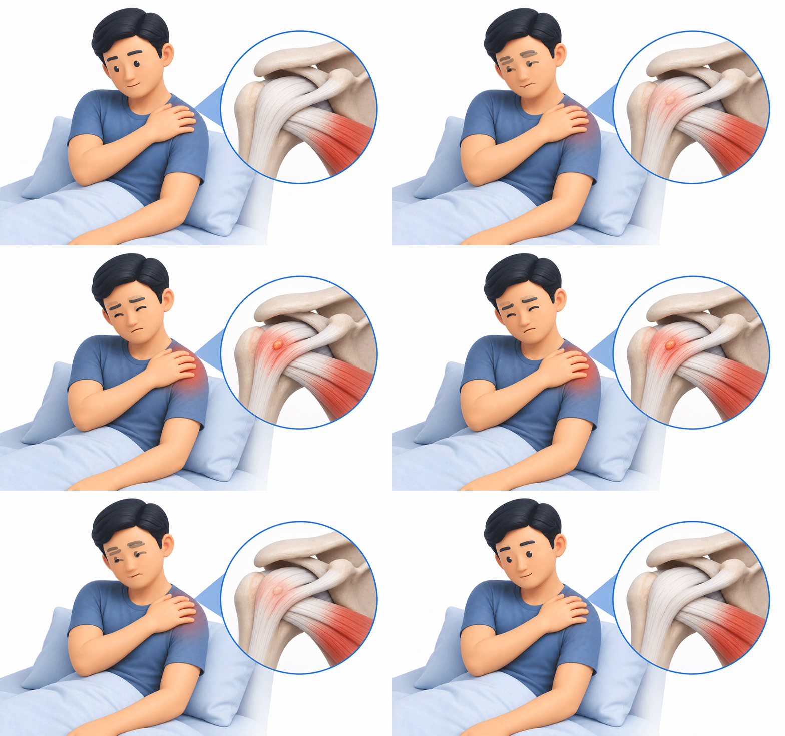
The inflammatory response that occurs as calcification in the shoulder tendon is resorbed may be closely related to pain that worsens at night.
Calcific tendinitis typically progresses through a formative phase (when calcium deposits build up), a resting phase (when the deposit remains clumped), and a resorptive phase (when it is gradually absorbed). During the resorptive phase, cells may recognize the calcium deposit as a foreign substance and begin clearing it, which can trigger a strong inflammatory reaction around the tendon.
At this stage, inflammation around the tendon and increased joint pressure can cause pain severe enough to disrupt sleep. A decrease in pain alone does not necessarily mean the calcification has completely disappeared.
2. What is the goal of the first non-surgical treatment?
In the initial stage of treatment, the goal is not to remove the calcification immediately, but to reduce pain and maintain shoulder range of motion.
When acute pain is severe, movement decreases and stiffness can remain in the joint. The general principle is to reduce pain through medication and activity modification, then continue rehabilitation to restore range of motion.
During periods of intense pain, rehabilitation intensity should be adjusted, but it’s important not to let the rest period become too long. If needed, a steroid injection into the subacromial bursa (beneath the acromion) may be considered. This injection is not intended to dissolve the calcification; rather, it is an adjunct option to reduce pain and create an opportunity to begin rehabilitation.
3. What should you look for in extracorporeal shock wave therapy (ESWT)?
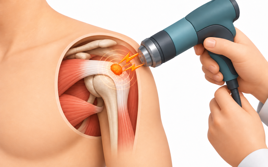
Extracorporeal shock wave therapy should be applied safely by considering both the type of energy used and the patient’s current disease status.
Studies suggest that high-energy focused ESWT tends to be more favorable than radial ESWT for pain relief, functional recovery, and reduction of calcification, although outcomes may vary depending on device performance. Focused ESWT concentrates energy on a single target point, while radial ESWT disperses energy over a broader area, so effectiveness can differ depending on the treatment goal.
If you are taking anticoagulants, inform your clinician before treatment due to bleeding risk. The number of sessions varies depending on the device, energy settings, and the patient’s symptom response, so it’s advisable to assess the effect after several sessions and then decide whether additional treatment is needed.
4. When do you choose barbotage?
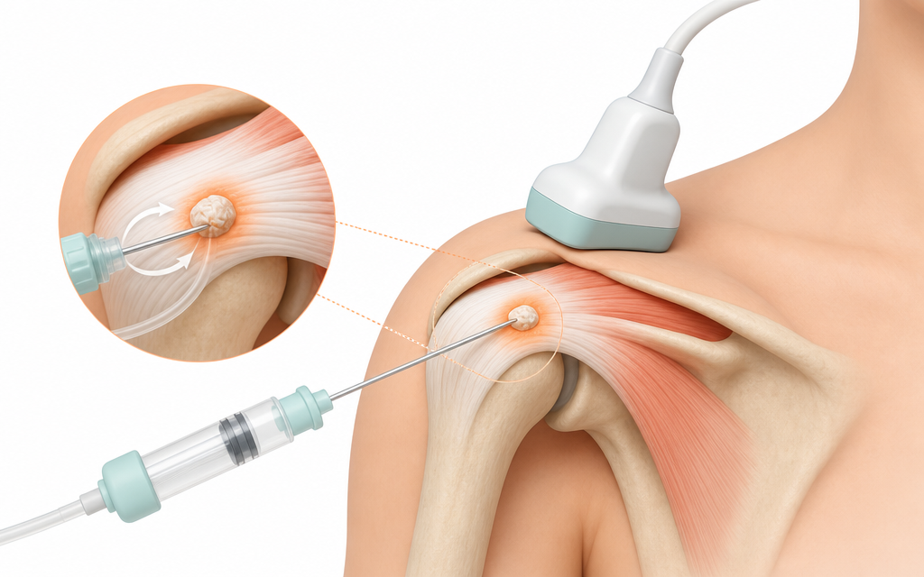
Ultrasound-guided barbotage is considered when impingement symptoms persist due to residual calcification, or when rapid pain control is needed over a short period.
This is a needle-based procedure in which the clinician visualizes the calcification with ultrasound, then irrigates it with normal saline and aspirates it. Because it involves direct needling, it may provide a strong response for early symptom control; however, the long-term calcification resorption rate may not differ substantially from standard injection-based treatment.
Because the procedure involves inserting a needle into the calcified area within the tendon, you should be aware in advance of possible temporary pain or minor bleeding immediately afterward. After the procedure, rehabilitation is needed to gradually stretch and restore shoulder mobility.
5. If there’s no pain, do you still need treatment?
If you have no pain and your daily function is maintained, it may be safer for joint health not to forcefully remove residual calcification.
Even if imaging shows some residual calcification after pain has subsided, there is no need to remove it if there are no additional symptoms. Some people feel anxious that leftover deposits might worsen symptoms and therefore want aggressive removal.
However, physically stimulating an asymptomatic deposit in the resting phase may increase the risk of micro-injury or inflammation in otherwise healthy tendon tissue. If there is no pain, it is preferable to track changes with periodic imaging and continue light rehabilitation as part of ongoing management. If pain or impingement symptoms develop later, the condition can be reassessed at that time.
6. Frequently Asked Questions (FAQ)
Q. How many ESWT sessions are usually needed?
The number of sessions depends on the device and energy settings used, the characteristics of the calcification, and the patient’s symptom response. After several sessions, clinicians typically decide whether to continue based on symptom sensitivity and tendon condition.
Q. Does ultrasound-guided barbotage work immediately after the procedure?
Because it involves needling and lavage, you may actually feel soreness or tightness immediately afterward. Symptoms may gradually improve over several days to weeks.
Q. Can a strong anti-inflammatory injection completely dissolve hard calcification?
A steroid injection only temporarily reduces inflammation; it does not directly dissolve and eliminate calcification. Repeated injections can affect surrounding tissues, so the number of injections must be carefully adjusted.
Q. If my arm movement is fine but calcification is seen, do I have to treat it?
If daily movement is normal and there is no pain, there is no medical need to force removal. It is appropriate to monitor symptoms periodically and reassess with imaging when necessary.
This health content has been reviewed by medical professionals, compiled from authoritative domestic and international sources, and is regularly updated in accordance with the latest clinical guidelines.
Sources
- de Witte PB, et al. Ultrasound-guided needling and lavage versus corticosteroid injection. Am J Sports Med. 2013.
- Louwerens JKG, et al. Extracorporeal shockwave therapy for calcific tendinitis. J Shoulder Elbow Surg. 2014.
- Rompe JD, et al. High-energy vs low-energy ESWT in chronic calcific tendinitis. J Shoulder Elbow Surg. 1998.
※ The copyright for all content on this blog belongs to medihi. Unauthorized copying, distribution, or derivative use is strictly prohibited, and violations may result in legal action without prior notice.