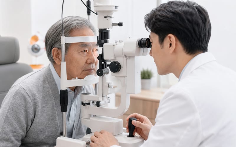

This post clearly explains what laser technology actually does in cataract & presbyopia surgery—and its medical limitations. For those who feel significant fear about the procedure, it also organizes practical criteria for choosing a safer surgical approach and an appropriate anesthesia plan.
“I’m really scared of eye surgery. Is there a way to do it without using a blade?”

The idea of having surgery while your eye is open can feel terrifying to anyone. Many people preparing for cataract surgery feel anxious about the thought of an instrument touching the eye, and advertisements often highlight appealing phrases like “bladeless, incision-free surgery.”
However, when considering cataract surgery, what clinicians evaluate first is not simply “whether a laser device is used.” The priority is whether your eye condition and the surgical plan fit together safely—and whether a highly anxious patient can tolerate the process as comfortably as possible.
If you read the medical criteria summarized below at your own pace, you can let go of vague fears and move toward a safer approach that fits you.
1. “Bladeless surgery”—does that mean no instruments are needed at all in the operating
room?
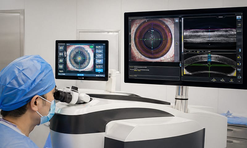
Laser-assisted cataract surgery (FLACS) is a method that uses a femtosecond laser to assist with certain steps of cataract surgery. A femtosecond laser is a highly precise laser that operates in extremely short pulses, and it can be used to create fine cuts or segment targeted tissue inside the eye.
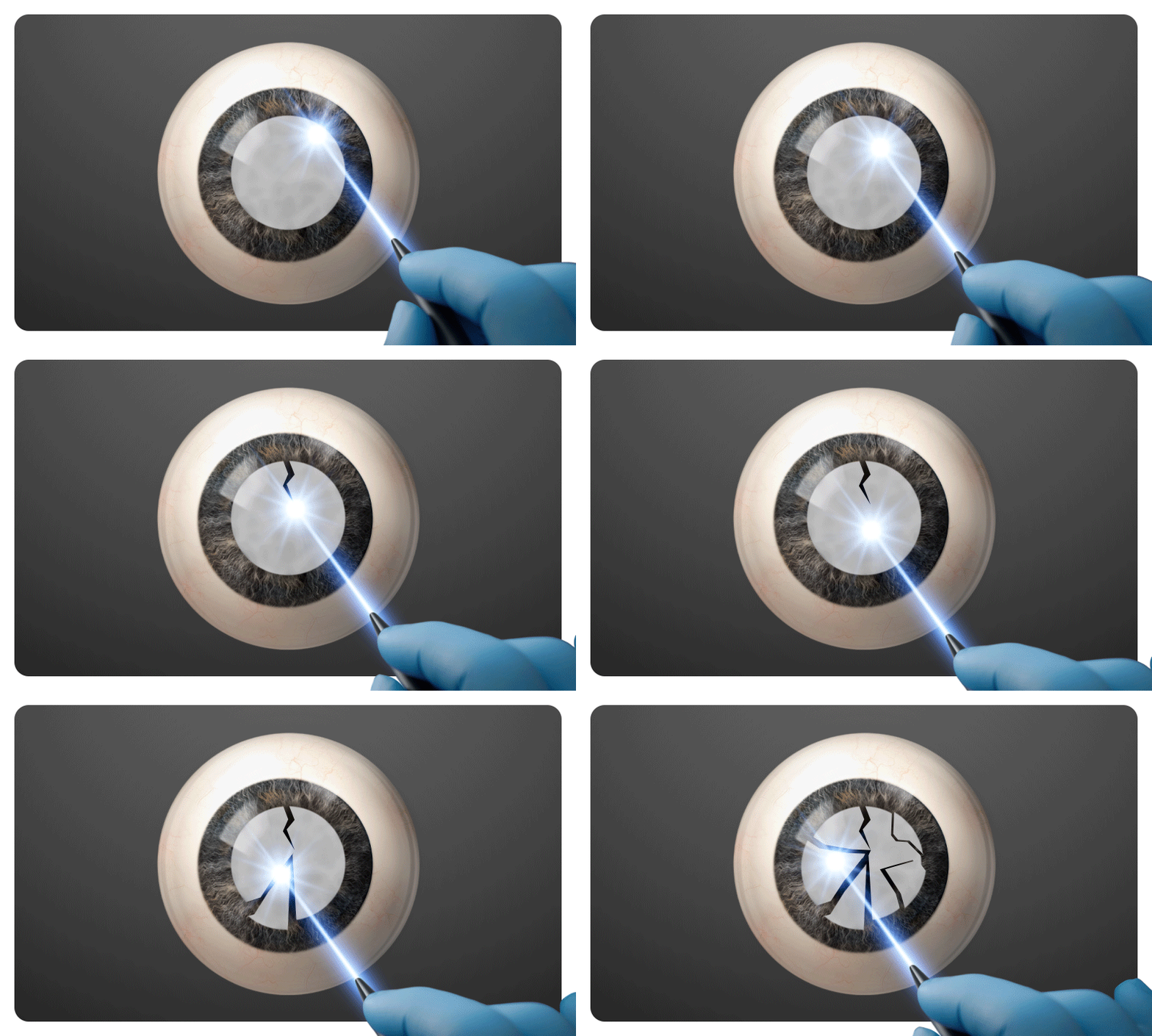
This is very similar to a semi-autonomous driving system in a car. The driver still leads the trip by understanding the overall route, while the system only helps in specific segments—such as lane keeping or speed control. Cataract surgery works in a similar way. Early in the procedure, the femtosecond laser can create tiny entry pathways in the cornea. It also operates only at specific steps, such as breaking up a hardened cataract into smaller pieces.
After that, the fragmented lens material must be safely aspirated. The key steps—placing a new intraocular lens (IOL) into the eye and completing the procedure—still require the surgeon’s direct hands-on work. So rather than saying “the laser performs the entire surgery,” it is medically more accurate to say that the laser assists certain steps that require high precision.
In fact, among ophthalmologists, cataract surgery is considered a technically demanding procedure that requires extensive experience and learning over time. For clinicians with less surgical experience, laser equipment can be a useful assistive tool for incision-making and fragmentation. But when an experienced specialist is operating, the laser-assisted steps may become an unnecessary added process from the patient’s perspective. The laser can be an excellent tool for precise cutting—but it cannot replace the entire surgical process.
2. When laser assistance may help reduce ultrasound energy use depending on your eye
condition
One major factor that affects recovery speed and corneal health after surgery is the amount of ultrasound energy used during the procedure. Cataract surgery removes the cloudy natural lens by breaking it up with ultrasound vibration and then extracting it. If ultrasound energy is used excessively, the corneal cells—meant to remain clear—can become stressed. As a result, corneal swelling right after surgery may last longer.
Think about how a semi-autonomous driving system can reduce engine strain on a steep mountain road. If the lens has become extremely hard—like a “mature” cataract—laser pre-fragmentation can help reduce the amount of ultrasound energy needed afterward to some extent. This can have a positive effect by reducing early postoperative corneal swelling.
On the other hand, if the cataract is not advanced and the lens remains relatively soft, standard ultrasound-based surgery alone can often be completed safely with low energy use.
Still, despite some potential benefits mentioned above, many large, reliable clinical studies report that laser-assisted methods do not provide a meaningful medical advantage over standard surgery in terms of final visual outcomes or prognosis. This is the mainstream view in ophthalmology worldwide. Therefore, the first step is to accurately assess your eye condition.
| Category | Situations Where Laser Method Can Be Helpful | Situations Where Standard Ultrasound Method Is Sufficient |
|---|---|---|
| Lens Condition | Mature cataract hardened like a stone | Mild, soft cataract with less progression |
| Intraoperative Indicators | Expected reduction in ultrasound energy use | Can be fragmented even with low ultrasound energy |
| Long-Term Visual Outcomes | No clinical difference between the two methods (proven by medical journals) | No clinical difference between the two methods (proven by medical journals) |
✅Checking which approach fits your eyes
- How hardened is your cataract at this stage?
- Is your case likely to require high ultrasound energy?
3. If you have strong fear of surgery, the “anesthesia plan” matters more than the device

Patients who feel vague but intense fear about eye surgery often start by searching the names of the newest devices. That’s because they hope a specific device will eliminate all discomfort. But the sense of stability a patient feels on the operating table depends more on anesthesia and sedation than on the type of equipment. Even the most advanced device is not helpful if you cannot relax.
To use a laser, a docking process is essential—this is when the patient’s eye must be precisely aligned and attached to the device. During this step, a heavy pressure sensation can occur as the eye is held in place. If someone has severe anxiety or claustrophobia, that pressure can intensify fear. And if a patient becomes so tense that they unconsciously move their head, it can interfere with the precision of the procedure.
There is also a medical risk that requires careful attention. During fixation, strong negative pressure is applied to the eye. Because cataract patients are often older, this can carry a risk of causing a peripheral retinal tear (break), and in severe cases may lead to retinal detachment.
Rather than insisting on a particular method, the priority should be an anesthesia plan that helps you stay calm. If eye-drop (topical) anesthesia alone feels difficult to tolerate, light sedation (sleep induction) may be considered. However, whether sedation is appropriate depends on your overall health, coexisting conditions, and the clinic’s treatment protocols. Discuss with your medical team to create a safe plan that fits your current condition. The less you move during surgery, the more safely the planned steps can be carried out.
4. Laser-assisted anterior capsule opening (capsulotomy)
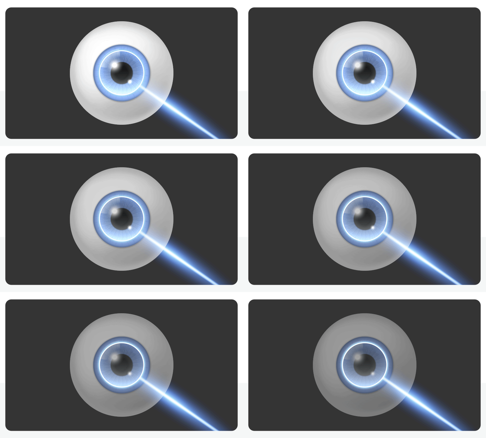
If you want to live more freely without glasses after surgery, you may consider a premium intraocular lens. This includes multifocal lenses and astigmatism-correcting lenses. To keep lens performance close to its intended target, it is crucial that the lens sits stably in the planned position inside the eye. This is where device precision can offer a specific advantage.
The step of cutting a round opening in the thin “bag” that surrounds the natural lens is called an anterior capsulotomy. This step is like laying the foundation that holds the intraocular lens steady. Some clinics may also suggest correcting astigmatism by making corneal relaxing cuts with a laser (Limbal Relaxing Incision, LRI). However, this method can leave subtle corneal haze or scarring that may cause glare, and astigmatism may recur over time as the incision heals.
For this reason, many well-known ophthalmologists worldwide increasingly prefer inserting a premium toric intraocular lens—during standard cataract surgery—to correct astigmatism in a more stable way, rather than relying on laser corneal incisions. Using such equipment is an additional out-of-pocket (non-covered) cost item. You should make a reasonable decision by considering your target vision level and the type of lens you need.
✅What to consider when choosing a lens
- Are you considering a premium lens that corrects astigmatism?
- Is capsulotomy precision likely to affect achieving your target vision with that lens?
5. Why vision can become foggy again after surgery
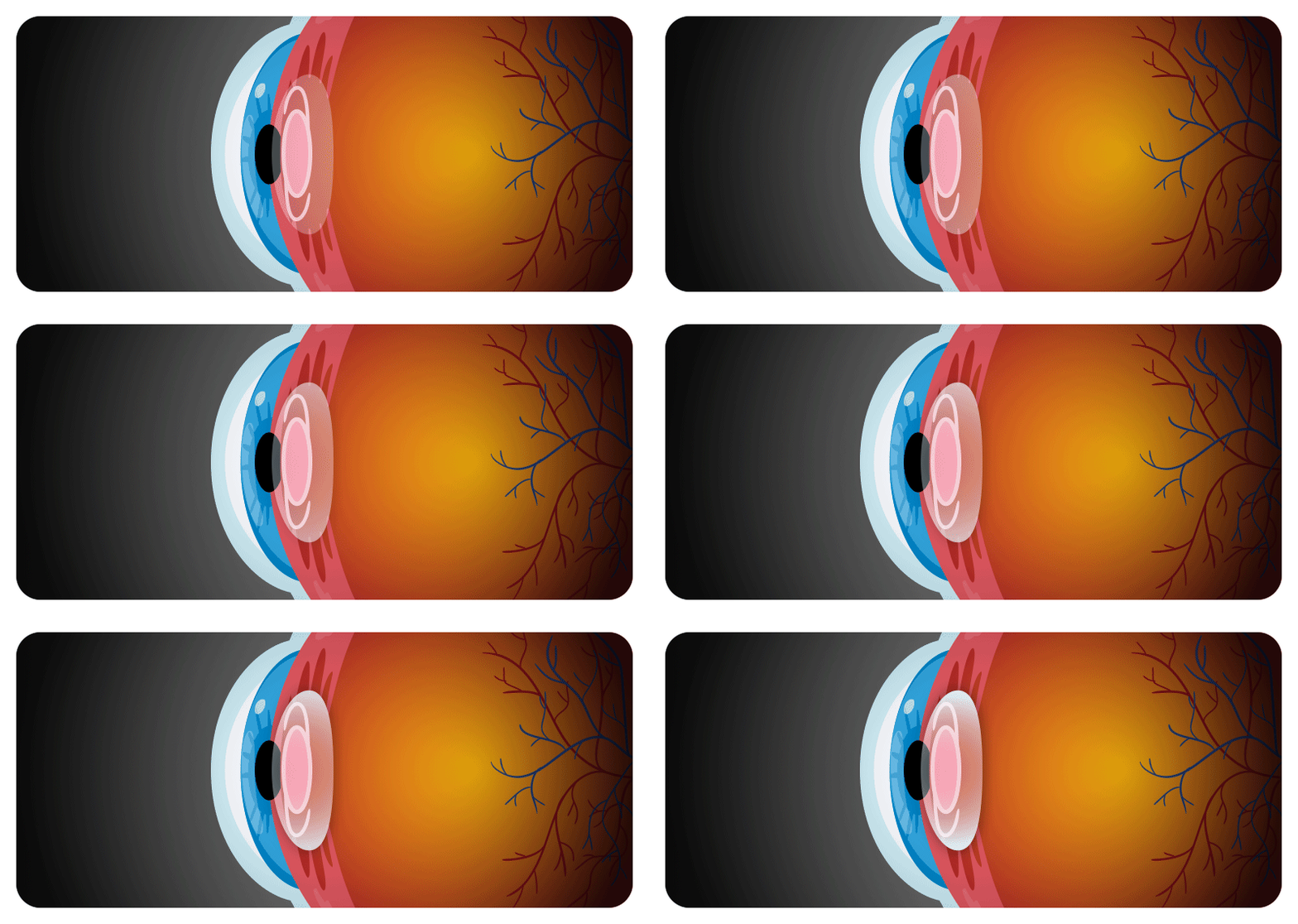
Some people notice their vision becoming cloudy again years after successful treatment. Many worry that something went wrong with the surgery. There is a reassuring medical fact: once the cloudy natural lens is completely removed, it does not grow back or “recur” in the body.
This phenomenon is medically called posterior capsular opacification (often referred to as a “secondary cataract”). It helps to imagine the eye’s structure as a “transparent cellophane pouch.” During cataract surgery, the back part of this pouch (the posterior capsule) is left in place to hold the intraocular lens securely. Over time, tiny residual cells can form a thin membrane on it, naturally causing the vision to look hazy.
Fortunately, fixing this is not as complex as the original surgery. It can be handled simply in an outpatient clinic using a YAG laser. The procedure creates a small opening in the cloudy back part of the “cellophane pouch” to clear the visual pathway. Once opened, it typically does not close on its own or become cloudy again, so a single treatment is usually enough to restore clear vision for the long term. If your vision becomes hazy after surgery, you do not need to panic—visit the clinic comfortably and have your condition checked.
6. Frequently Asked Questions (FAQ)
Q. How exactly is laser-assisted cataract surgery different from the conventional method?
In the standard method, the surgeon directly creates an entry through the cornea with fine instruments and breaks up the lens. In the laser-assisted method, the device performs some of these steps. However, many clinical studies published in international journals have shown that the laser method has no statistical advantage over standard surgery, and many prominent U.S. ophthalmologists are also trending toward choosing standard surgery over
laser.
Q. I’m worried the pain during surgery will be severe. How is anesthesia usually done?
Typically, topical anesthesia is provided using anesthetic eye drops, so sharp pain is uncommon. However, when using laser equipment, the step that fixes the eye with strong negative pressure can cause a heavy pressure sensation. In older patients, there is a rare concern for side effects such as peripheral retinal tears, so if your retina is fragile you should discuss this in advance with your clinician. If fear is severe, sleep induction (sedation anesthesia) may be added.
Q. Does using laser technology make recovery faster than standard surgery?
In patients with very hard cataracts, laser pre-fragmentation may reduce ultrasound energy use and lead to less early corneal swelling. However, many studies report that long-term final visual recovery outcomes are generally comparable to the standard method. Recovery speed varies by individual depending on retinal condition and how well inflammation is managed.
Q. My vision doesn’t feel like it used to. When is it safest to get an eye consultation?
Rather than focusing only on a vision number, the key is how much daily life is affected. If reading becomes difficult or nighttime driving causes fatigue due to glare, it’s a good time to start a consultation. If light sensitivity worsens or you see double images, it’s recommended not to delay an exam.

The most confusing part before cataract surgery is likely staying grounded amid the flood of information. As you can see after reading this far, what determines satisfaction is not simply the most expensive or newest device. More important factors are how advanced your cataract is, what type of lens you need, and whether you can tolerate the surgical environment comfortably.
I hope you won’t be led by vague fear into relying too heavily on device names alone. Based on the objective criteria reviewed today, have a deeper conversation with your clinician. If you build the most suitable treatment plan for your eyes in a psychologically comfortable state, you can safely welcome a clearer, cleaner field of vision.
Sources
- The Korean Ophthalmological Society: General patient guidance on cataract diagnosis and treatment
- Health Insurance Review & Assessment Service (HIRA): General guidance on DRG (Diagnosis-Related Group) cataract surgery
- Summaries of comparative outcome analyses between phacoemulsification and FLACS, including the FACT Trial (randomized trial)
※ The copyright for all content on this blog belongs to medihi. Unauthorized copying, distribution, or derivative use is strictly prohibited, and violations may result in legal action without prior notice.
Recommended reads
 medihi