

We’re taking a closer look at traumatic cataract that can occur after a strong blow to the eye during sports or everyday activities. It’s not enough to confirm “just a cataract”—it’s essential to carefully check whether other nerves or blood vessels inside the eye have also been injured, and then determine the safest timing for surgery based on the individual eye condition. We also summarize long-term care strategies and emergency response tips you should know to protect your vision after surgery.
"During sports, I got hit hard around the eye, and now my vision in one eye suddenly looks blurry."

After a strong impact to the eye during sports or daily life, many people feel alarmed when their vision suddenly worsens. Unlike age-related cataracts that develop gradually over time, a cataract caused by injury can appear abruptly right after the trauma.
When you visit the clinic for an eye injury, an ophthalmologist does not only check whether a cataract has formed. The most important step is to assess the overall health of the eye—especially whether internal nerves (the retina) or other structures have been injured as well. Below, we explain in a clear and practical way the key principles and treatment process you should know to treat the injured eye safely and recover vision.
1. If your vision drops after an eye injury, never brush it off
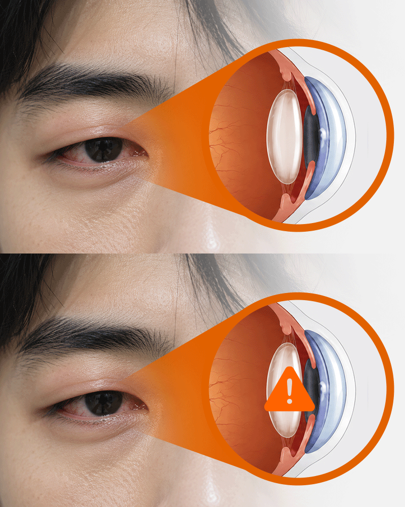
When the eye receives a blunt, strong impact, the visible external injury does not always reflect what is happening inside. Even if it looks mild—such as slight redness or eyelid swelling—you should still consider the possibility of a sudden change in intraocular pressure or damage to internal structures.
A strong external impact can cause the normally clear lens (which functions like a camera lens) to turn white and cloudy—this is called a traumatic cataract. However, it can be risky to assume that reduced vision after trauma is caused by “just a cataract.” Multiple serious problems may be involved, such as intraocular bleeding, elevated intraocular pressure, retinal damage, or optic nerve injury.
At the hospital, one of the first things checked is whether there is any tearing or puncture of the eye surface—an open globe injury. If the white part (sclera) or the clear front surface (cornea) is torn, the pressure inside the eye can drop sharply and internal contents may prolapse outward, creating an emergency. Because this cannot be judged by simple redness alone, a detailed examination is essential.
If there is even a small possibility that the eye is torn, you must not rub or press on it. Pressure can force internal structures out of the eye, creating a highly dangerous situation. Immediately after injury, protect the eye from pressure using a rigid eye shield or even a plastic cup, and seek ophthalmic care as soon as possible for precise testing such as intraocular pressure measurement and retinal/nerve evaluation.
2. Vision loss after trauma may not be a cataract: why checking associated injuries
matters
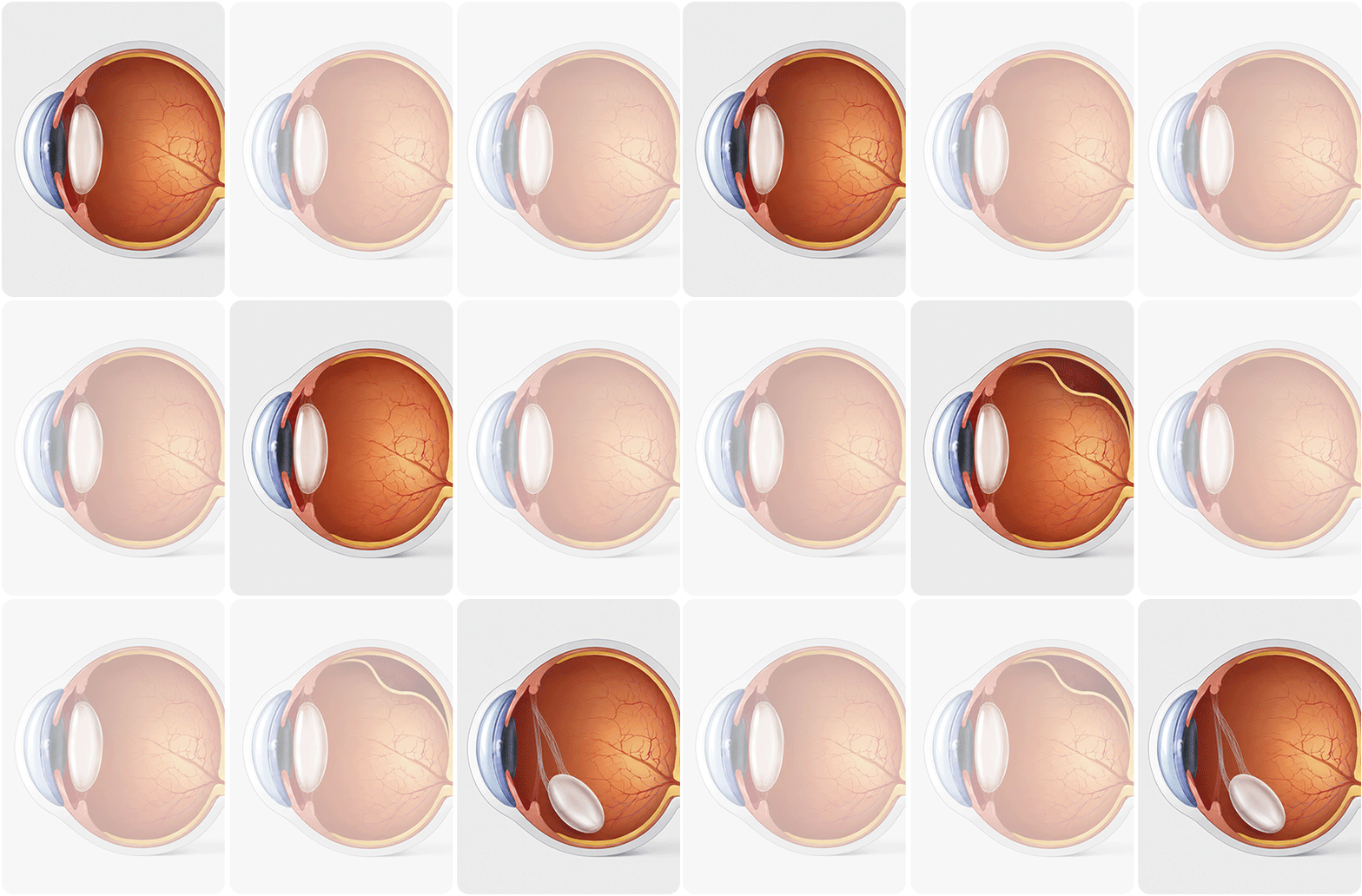
When vision drops after an impact, it’s easy to think, “I must have developed a cataract.” But in many cases, what determines how well vision will recover later is not the cataract itself, but whether other internal eye structures were injured at the same time.
A common example is intraocular bleeding (hyphema). When bleeding occurs inside the eye, the drainage pathway for the eye’s fluid (aqueous outflow) can become blocked by clots, causing intraocular pressure to rise rapidly. That’s why it’s crucial to check whether blood is pooling and whether pressure is increasing.
Retinal injury also requires great caution. A strong impact can tear the retina (retinal tear) or cause it to detach from the eye wall (retinal detachment), which is a serious condition. If the central area responsible for detailed vision is affected, vision recovery can be limited even after surgery. If bleeding is severe or the cataract prevents a clear view of the back of the eye, an ultrasound exam is often needed to carefully check for hidden retinal damage.
In addition, the thin fibers (zonules) that hold the lens in place can break due to trauma. When this happens, the lens can shift out of position (lens dislocation), leading to symptoms such as double vision or severe visual distortion. For these reasons, removing the cloudy lens alone may not immediately restore vision in an
injured eye. During evaluation, an ophthalmologist systematically checks for associated injuries and builds a safe treatment plan.
✅Checklist of associated injuries doctors must evaluate in traumatic cataract care
- Intraocular bleeding and re-bleeding: checks whether clots are raising intraocular pressure or damaging the cornea.
- Lens dislocation: checks whether broken zonules are allowing the lens to shift or wobble out of position.
- Elevated intraocular pressure and traumatic glaucoma: monitors whether damage to the drainage pathway is raising pressure and compressing the optic nerve.
- Retinal and posterior segment injury: confirms via detailed exam and ultrasound whether the retina has torn or detached.
- Optic nerve injury: evaluates whether the optic nerve—the key pathway connecting the eye and brain—was directly affected.
3. Early surgery vs observation: how surgical timing is decided in traumatic cataract
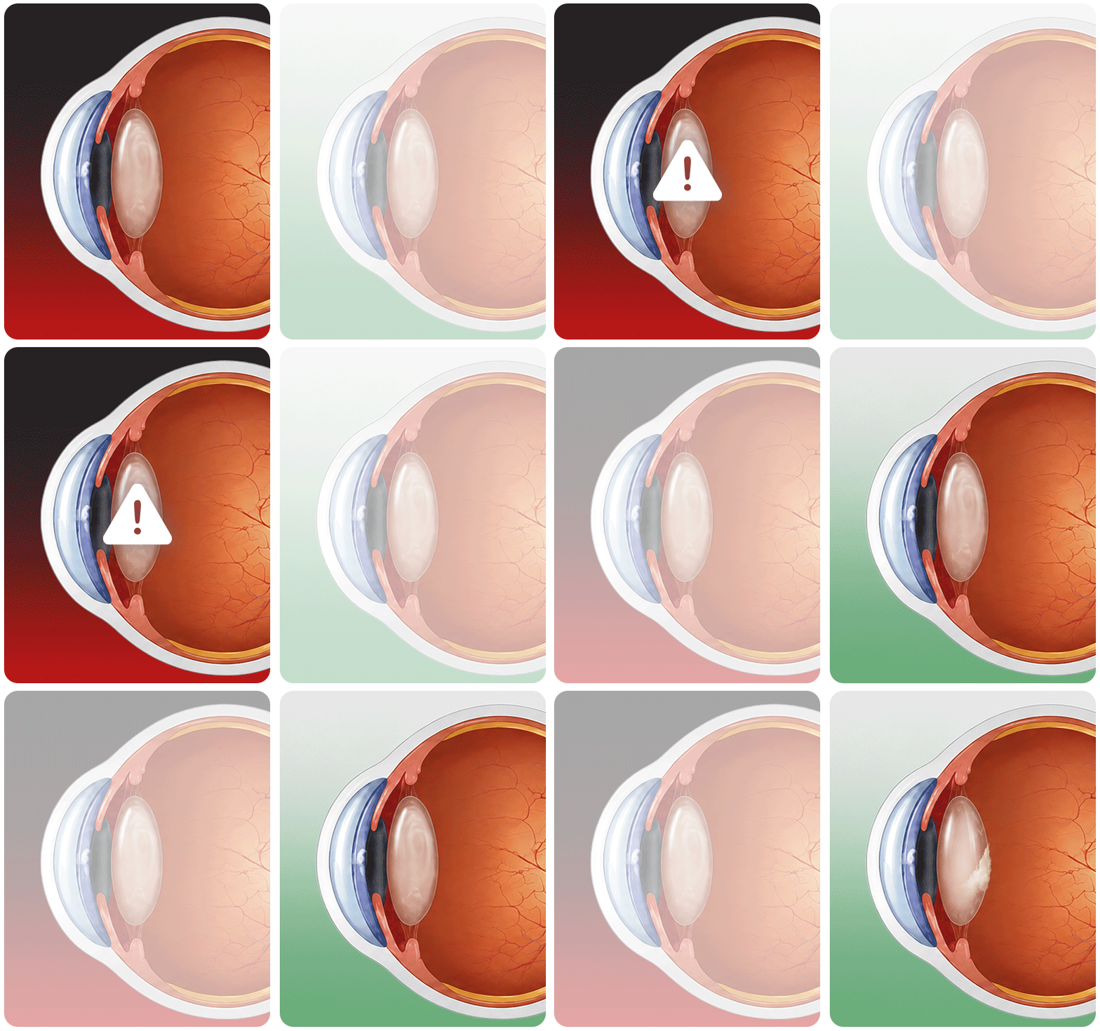
The timing of surgery for traumatic cataract cannot be decided using the same criteria as typical age-related cataracts. In routine cataracts, the main factor is how much the patient feels limited by blurry vision in daily life. In traumatic cases, however, the decision must be made after carefully assessing the overall eye condition—such as the degree of inflammation, changes in intraocular pressure, whether the lens capsule has ruptured, and how much other internal tissue has been injured.
Most importantly, there are emergency situations where prompt surgery is necessary. A key example is rupture of the “capsule” (lens capsule) that surrounds the lens. If the capsule tears, internal lens material can leak into the eye and trigger severe inflammation or acute glaucoma, requiring urgent treatment. Surgery may also need to be expedited if the injured lens swells and blocks fluid flow, causing a rapid pressure spike, or if the lens shifts forward and risks damaging the inner surface of the cornea.
On the other hand, there are cases where it is reasonable to treat with medication, observe, and schedule surgery more safely. If the lens capsule is intact, the cataract is limited to part of the lens, intraocular pressure remains stable, and associated injuries are mild, there may be no need to rush.
| Situations (Signals) Requiring Early Surgical Intervention | Conditions Allowing for Planned Surgery and Monitoring |
|---|---|
| Lens capsule rupture | Intact lens capsule, localized opacity |
| Risk of uveitis or glaucoma due to lens protein leakage | Intraocular pressure (IOP) stably maintained within normal range |
| Increased intraocular pressure due to lens intumescence | Mild accompanying injuries, allowing for observation |
| Progressive corneal endothelial damage caused by anterior dislocation | Inflammatory response controlled by medication |
Immediately after a severe eye injury, internal tissues often swell and inflammation becomes active. Operating during this highly sensitive period can further damage already weakened structures such as the zonules or capsule. Therefore, unless emergency surgery is required, the general principle is to calm the eye first with anti-inflammatory drops and pressure-lowering medication, then proceed with surgery at the safest time.
4. Long-term care after surgery: how to protect your vision over time

Even if cataract surgery goes smoothly, treatment of an injured eye is not necessarily “finished.” After a major impact, delayed complications can appear months or even years later, so consistent follow-up is essential.
A representative late complication is traumatic glaucoma. If the drainage pathway for aqueous fluid was subtly damaged at the time of injury, it may gradually become obstructed over time, raising intraocular pressure. Because this can damage the optic nerve without noticeable symptoms early on, regular pressure checks remain necessary even after surgery.
Another concern is intraocular lens (IOL) instability or dislocation. If the zonules that support the implanted lens were already weakened by the trauma, the lens can shift or dislocate years later. If you suddenly experience double vision or severe glare, you should return for evaluation of lens position.
In addition, previously hidden retinal problems may be discovered later. Small retinal tears or swelling that were not visible immediately after injury can become apparent over time.
So even if your vision seems good after surgery, you should continue to review the following items regularly with your doctor.
✅Postoperative checklist to review with your doctor
- Changes in your visual field: compare how vision feels now versus immediately after the injury.
- Presence of additional symptoms: check whether flashes of light (photopsia), increased floaters, severe glare, or pain have appeared.
- Regular follow-up: confirm that intraocular pressure is stable, the optic nerve is healthy, and the IOL remains well positioned.
- Timing of return to sports: decide step-by-step—based on objective medical assessment, not personal feeling—when to resume everything from light daily activity to high-impact sports with risk of collision or ball strikes.
5. How does treatment differ between adults and children?
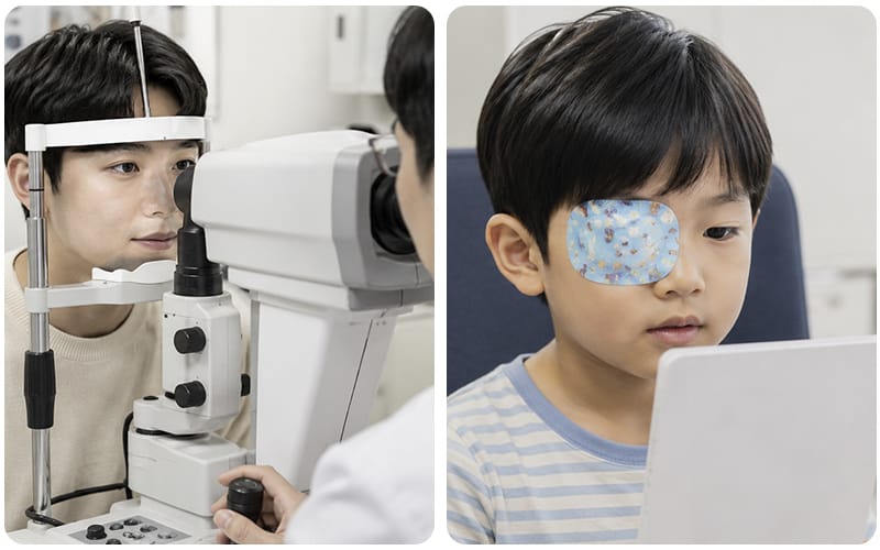
When treating a cataract caused by trauma, the goals of care and postoperative
management differ greatly depending on whether the patient is an adult or a child. This is because in children, the most important consideration is how the eye and brain’s visual pathways develop (the critical period of visual development).
For adults, visual development is already complete. The main goal is to restore vision safely and return to work and daily life. After surgery, clinicians monitor whether intraocular pressure rises and whether the lens remains stable, and they coordinate the timing of returning to activities.
For children, the approach is fundamentally different. From birth until around age 8, children go through a critical period when vision and brain pathways are actively developing. If a cataract blocks clear vision during this time, visual stimulation may not reach the brain properly, and a long-lasting visual impairment called amblyopia (“lazy eye”) can develop—even with glasses.
Therefore, even after surgically removing the cloudy lens, children require much more meticulous care to support normal visual development. Children are also more prone than adults to the back of the lens area becoming cloudy again, so additional preventive steps are often performed during the initial surgery to reduce the chance that the visual axis becomes blocked again.
Postoperative management is also crucial. Vision should be corrected promptly with glasses or contact lenses, and patching therapy may be used—covering the healthy eye for certain periods to encourage use of the injured eye. The schedule and duration of patching are adjusted based on the child’s age and vision status, and consistent follow-up and treatment adherence by caregivers can strongly influence the child’s lifelong vision.
6.Frequently Asked Questions(FAQ)
Q. Right after the injury I couldn’t see well, but now my vision seems back to normal. Do I still need an eye exam?
Yes. Even if symptoms seem to improve naturally, you should still undergo a detailed examination. After a strong impact, vision can temporarily drop and then recover as the inside of the eye “bruises” and settles. However, even without pain or obvious symptoms, there may be a tiny retinal tear or damage to the drainage pathway that causes intraocular pressure to rise gradually. These issues can progress quietly and later lead to major vision
loss from conditions such as retinal detachment or glaucoma—so you should be checked even if you feel better.
Q. What criteria determine the timing of surgery for traumatic cataract?
Surgical timing is not decided based only on how blurry you feel your vision is. Doctors evaluate the overall health of the eye, including whether the lens capsule is ruptured, whether the fibers supporting the lens are broken and the lens is unstable, the degree of inflammation and intraocular pressure, and whether the posterior structures (retina and optic nerve) are intact. Based on this, they decide whether emergency surgery is needed immediately or whether it is safer to calm the eye with medication first and plan surgery at a time with fewer risks.
Q. How long does it take before I can return to sports after surgery?
The timing varies widely depending on how severe the injury was and how extensive the surgery was. Light daily activities such as walking can often be resumed relatively early. However, you should not restart high-impact sports—such as ball sports or combat sports—based only on how you feel. The principle is to increase activity step-by-step only after your doctor objectively confirms that the implanted lens is stable and that intraocular pressure and the retina are safe.
Q. After an eye injury, what symptoms mean I should go to the ER (ophthalmology) right away?
If you develop any of the following symptoms after injury, you should seek emergency care without delay.
- A sudden, sharp drop in vision
- Severe eye pain and headache that do not improve even after taking pain medication
- Flashes of light, or a sudden increase in floaters (specks or “gnats” drifting in vision)
- Part of your visual field becoming blocked, like a black curtain
These can be warning signs of conditions requiring immediate treatment, such as retinal detachment or a rapid rise in intraocular pressure. How quickly you get to the hospital can significantly affect visual recovery, so you should come in as soon as you notice them.

We reviewed the clinical approach to decreased vision following blunt ocular trauma. Traumatic cataract should not be approached as a single lesion of lens opacity alone; systematically evaluating associated intraocular injuries—such as hyphema, zonular damage, retinal detachment, and traumatic optic neuropathy—is central to determining visual prognosis.
Deciding the timing of surgery should also be based not on subjective discomfort, but on an integrated assessment of lens capsule integrity, intraocular pressure trends, intraocular inflammatory response, and the extent of associated posterior segment injury. Performing surgery once ocular tissues have sufficiently stabilized is a key principle for minimizing intraoperative complications and optimizing outcomes.
Even after successful completion of surgery, long-term follow-up must continue to monitor for delayed complications such as traumatic glaucoma, late IOL dislocation, and delayed onset of retinal pathology. Maintaining a regular surveillance plan—including intraocular pressure, optic nerve status, IOL position, and retinal evaluation—is a practical requirement for preserving long-term visual function.
Accurately identifying the full spectrum of intraocular injury at the initial assessment and building an individualized treatment plan based on that evaluation is the starting point of an evidence-based approach aimed at achieving optimal visual recovery in patients with traumatic cataract.
Sources
- Korea Disease Control and Prevention Agency (KDCA) National Health Information Portal, Eye trauma (traumatic cataract, hyphema, traumatic retinal detachment)
- Journal of the Korean Ophthalmological Society (JKOS), Incidence and risk factors of intraocular lens dislocation in Korea (claims-data–based study)
- International Ophthalmology, Visual outcomes of open globe injury patients with traumatic cataracts (2021)
※ The copyright for all content on this blog belongs to medihi. Unauthorized copying, distribution, or derivative use is strictly prohibited, and violations may result in legal action without prior notice.
Recommended reads
 medihi