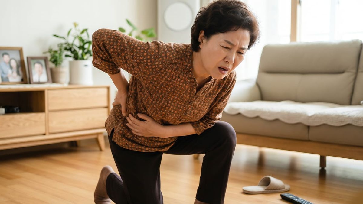

Lumbar disc herniation (lumbar herniated disc) and lumbar spinal stenosis can look and feel similar because both may cause low back pain and leg symptoms. However, what makes symptoms worse—and which positions bring relief—often differs. This post organizes common pain patterns using three practical criteria: walking, posture, and pain radiating into the leg, and reviews when diagnostic testing (such as MRI) becomes more helpful.
“My back hurts—does this mean it’s a disc problem, or stenosis?”
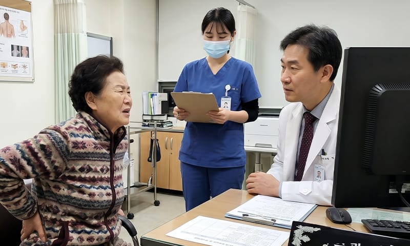
Many people visit for low back pain and ask what their diagnosis is. In the exam room, the first priority is not the label itself. Clinicians first assess whether a nerve is truly being compressed and whether those neurological signs appear to be progressing. Even with the same “back pain,” priorities can differ depending on whether the main issue is nerve root irritation or narrowing of the spinal canal.
When pain persists, it’s common to think about imaging tests first. But the starting point for distinguishing causes is often a simple daily record: noting when pain turns “on” and “off,” and what triggers or relieves it. Most spine-related pain can be evaluated step by step, and in many cases it’s appropriate to begin with a structured approach before rushing into advanced testing.
1. Lumbar disc herniation vs. lumbar spinal stenosis: differences in how pain occurs
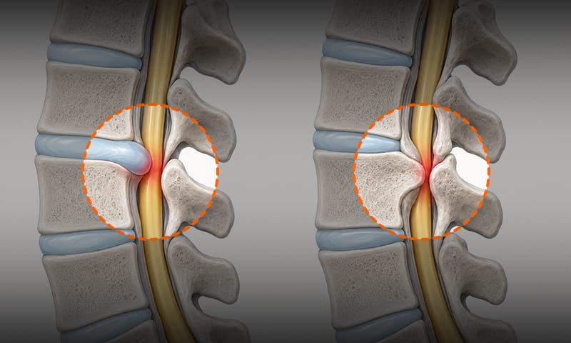
Inside the spine is a passageway called the spinal canal, where nerves travel. If you imagine it as a “tunnel,” it becomes easier to understand. Lumbar spinal stenosis is a degenerative process in which bone, facet joints, and ligaments thicken over time, gradually narrowing the tunnel itself. As the overall pathway for the nerves becomes tighter, the nerves can be compressed. That’s why many people report gradually increasing discomfort starting in their 50s and beyond.
In contrast, lumbar disc herniation occurs when the disc material that normally acts like a cushion (the nucleus pulposus) shifts out of place. That displaced disc material can irritate a specific nerve root, sometimes with true compression as well. This is why some people feel their pain worsened relatively suddenly.
On imaging, nerve compression can appear in both conditions. However, treatment is not determined by a single scan. Imaging findings become clinically meaningful when they match the symptom pattern and physical examination.
- Lumbar disc herniation: displaced disc material irritates a nerve root
- Spinal stenosis: the “tunnel” the nerves travel through gradually narrows
- Shared point: imaging alone may not definitively identify the main pain generator
2. How to tell them apart by walking and posture
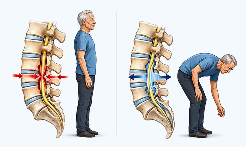
A classic pattern often discussed in spinal stenosis is neurogenic intermittent claudication (leg pain or heaviness that becomes so severe after walking a short distance that you must stop and rest). Because the problem is nerve-related, walking or standing can trigger leg numbness, tingling, or pain, but symptoms repeatedly improve with sitting or bending forward.
Think of the nerve tunnel as a narrow road. When you stand or walk, the lower back extends and the tunnel can become tighter—like traffic getting blocked. When you sit or bend forward, there is relatively more space. As the “blocked road” opens up, pressure on the nerves can decrease.
However, exceptions exist, so “bending = disc” and “extending = stenosis” should not be treated as a fixed rule. Posture-related changes are best used as practical clues to share with your clinician.
✅ Self-check checklist for walking/posture patterns
- After you start walking, at what point (minutes/meters) do you have to stop?
- Once you stop, how long does it take before you can walk again?
- When you sit, do you feel better right away, or does it take time?
- If you lean slightly forward while holding a shopping cart, does walking become easier?
3. Pain radiating down one leg: a clue that suggests a disc problem
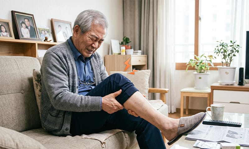
A frequently observed clue in lumbar disc herniation is one-sided radicular pain (radiating pain down one leg). Pain may travel from the buttock to the thigh, calf, and foot, and numbness or an “electric,” shooting sensation may occur as well. This pattern often becomes more distinct when displaced disc material strongly irritates a specific nerve root.
Many people also feel that discomfort worsened relatively suddenly. Coughing or sneezing can sometimes send pain “shooting” into the lower back and down the leg.
However, in middle-aged adults (50s–60s), there are many variables. Disc herniation and spinal stenosis can also occur together, so it’s difficult to conclude the cause based only on sudden onset. It’s important—together with a clinician—to check for areas of reduced sensation or new weakness and then narrow down the primary driver.
4. Is an MRI always necessary? Timing of tests and warning signs

MRI is a helpful, detailed test. But it does not always need to be done immediately when symptoms begin. Many guidelines consider MRI most meaningful when it will change the treatment plan—such as decisions about surgery or interventional procedures—because disc bulging or stenosis can also appear on imaging in people without symptoms.
That’s why clinicians first ask about daily patterns: when it hurts, how far you can walk, and whether symptoms change with posture. They then check for signs of nerve root involvement on physical examination before deciding whether imaging is needed.
That said, there are warning signs (“red flags”) that require prompt evaluation. Progressive muscle weakness is a key example. Signs can include repeatedly losing a slipper without noticing, or frequently tripping on flat ground because the ankle cannot dorsiflex well (foot drop). Urgent assessment is also needed if bowel/bladder dysfunction or reduced sensation in the perineal (“saddle”) area occurs. In these situations, evaluation at a medical facility without delay is recommended.
✅ Red-flag checklist: when care is urgent
- Is leg strength gradually getting worse? (Check whether one side is worse.)
- Are slippers slipping off easily, or is it hard to lift the front of the foot?
- Has bowel/bladder sensation become unusually dull compared with normal?
- Has sensation decreased around the perineum or buttock area?
5. Medications, exercise, acupuncture, surgery: how to choose step-by-step treatment

In most cases, treatment prioritizes non-surgical approaches and encourages maintaining activity rather than stopping completely. Exercise and rehabilitation are recommended within the limits allowed by pain, because prolonged bed rest can reduce muscle strength and slow recovery.
Korean medicine treatment, as one form of conservative care, may help with short-term pain control and reducing inflammation around the nerve.
- Pharmacopuncture and bee venom therapy: In addition to conventional injection-based treatments, inflammation and swelling related to nerve irritation may be reduced by injecting pharmacopuncture (refined herbal extracts) or bee venom near the target nerve root to help calm symptoms more quickly.
- Chuna manual therapy: The narrowed spinal passage in stenosis and the uneven mechanical loading seen with lumbar disc herniation may be addressed through Chuna manual therapy, in which a Korean medicine doctor manually adjusts spinal joints to help create more space and reduce nerve compression.
- Herbal medicine: Recovery of irritated nerves and strengthening of ligaments around the spine may be supported through individualized herbal prescriptions aimed at promoting functional and structural improvement.
Surgery is considered when functional decline remains significant despite conservative care, or when neurological deficits—such as clear, progressive weakness—are present. If medications, exercise, and injections are ways to “make a blocked road less blocked” or “use detours better,” surgery is an option to “widen the road” or “remove what is pressing on the nerve.” Key criteria include whether signs of nerve compression are progressing and whether functional decline is substantially interfering with daily life.
6. Frequently Asked Questions (FAQ)
Q. My legs get numb whenever I walk—does that mean it’s a disc problem or stenosis?
If symptoms worsen with walking or standing and improve with sitting or bending forward, this may resemble the neurogenic claudication pattern seen in lumbar spinal stenosis. On the other hand, if—regardless of walking distance—bending forward triggers sharp, electric-like pain radiating down one leg, lumbar disc herniation may be suspected. For accurate differentiation, a physical examination is recommended.
Q. Why do I feel okay when sitting, but it hurts when I stand?
In spinal stenosis, the nerve passageway is already narrowed. Standing or walking can extend the lower back and narrow the passage further, making symptoms more noticeable. Conversely, sitting or bending forward can relatively widen the passage, reducing nerve pressure and easing pain.
Q. My MRI shows a disc problem—do I have to get surgery?
Surgery is not decided based on imaging alone. A structural finding may not be the only cause of pain. Surgery is considered when there is no improvement with conservative treatment and functional decline is significant, or when warning signs such as muscle weakness are present.
Q. When should I seek medical care right away?
Evaluation is recommended if leg strength is progressively decreasing—for example, frequent tripping because the ankle or toes are weak. If bowel/bladder dysfunction, reduced perineal sensation, or other warning signs such as fever, a history of cancer, or trauma are present, you should be examined by a clinician without delay.
When you’re unsure what’s causing your low back pain, it’s often more helpful to set practical criteria rather than deciding on a diagnosis first. Observe whether symptoms worsen with walking or standing, and whether they improve with sitting or bending forward. Whether pain radiates down one leg is also an important clue.
If you record your pain patterns, discussions with clinicians become much easier. If there are no red flags, a step-by-step, non-surgical approach is commonly recommended. The goal is to understand what your body is signaling and return to daily life safely using reasonable, evidence-informed criteria.
Sources
- Kim et al., Comparison of caudal epidural injection and selective nerve root block in LDH or stenosis, J Korean Orthop Assoc (2022)
- Jin et al., A systematic review of treatment guidelines for lumbar disc herniation, Neurospine (2025)
- WHO Low back pain fact sheet (2023)
※ The copyright for all content on this blog belongs to medihi. Unauthorized copying, distribution, or derivative use is strictly prohibited, and violations may result in legal action without prior notice.
Recommended reads
 medihi